 The University of Chicago Digestive Diseases Review 2017 (Videos+PDFs)
  MKSAP® 19 Audio Companion Part B
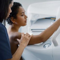
Designed for both the general and specialized radiologist, UCSF Abdominal and Thoracic Imaging is an online CME course providing an extensive review of clinically relevant topics in chest, abdominal, pelvic, and ob-gyn imaging. Speakers discuss interpretation tips for both routine and emerging applications, artificial intelligence and its implications in clinical practice, and more.
You get case-based continuing medical education lectures targeting relevant areas of focus, including:
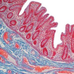
- Abdominal — female pelvis, acute pelvic pain in the reproductive age female, early and ectopic pregnancy, male pelvis and prostate, gastrointestinal conditions including the liver, and organ transplants
- Thoracic — lung cancer screening, pulmonary embolism, mediastinal masses, chest radiographs, and other findings and indications relevant for the general radiologist
Learning Objectives
At the conclusion of this activity, the participant will be able to:
- Recognize complications and diagnostic approach to a pregnant patient, including first trimester viability and placenta accrete spectrum disorder
- Distinguish between benign and malignant conditions of the female pelvis
- Identify and evaluate abdomino-pelvic conditions relevant to the prostate, bowel, uterus, biliary tree, and abdominal transplants
- Valuate acute pelvic pain in both the pregnant and non-pregnant female
- Implement improved thyroid biopsy procedural skills
- Differentiate between typical and atypical appearances of thoracic emergencies and pulmonary infections
- Identify pathologic conditions of benign incidentalomas and mimics in the lungs, heart, and mediastinum
- Implement a multidisciplinary approach to the diagnosis of lung cancer, including lung biopsy techniques
- Utilize and understand the basis and implications of artificial intelligence in clinical practice
- Understand the fundamentals of developing imaging techniques and CT/MR safety
Intended Audience
Radiologists and other medical professionals who will benefit from a greater understanding of image interpretation and diagnosis.
TOPICS / SPEAKERS
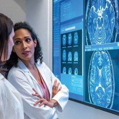
Female Pelvis and Pregnancy
MRI Safety – Michael A. Ohliger, MD, PhD
MRI of Gynecologic Malignancy – Michael A. Ohliger, MD, PhD
Practical Approach to Adnexal Imaging – Liina Poder, MD
MRI in Pregnancy – Non-Fetal Indications – Liina Poder, MD
Acute Pelvic Pain – Positive Pregnancy Test – Liina Poder, MD
Acute Pelvic Pain – Negative Pregnancy Test – Liina Poder, MD
Abdominal-Pelvic Q&A – Clinical Scenarios – I – Michael A. Ohliger, MD, PhD and Liina Poder, MD
Abdominal & Pelvic Imaging
Prostate T2 and Diffusion – Technical Tips, Pearls and Pitfalls – Michael A. Ohliger, MD, PhD
Imaging Mimics of Gynecologic Malignancy – Michael A. Ohliger, MD, PhD
Pearls and Pitfalls in Biliary MRI – Michael A. Ohliger, MD, PhD
MRI for Endometriosis – Value-Added – Liina Poder, MD
Beyond Torsion and Infection – Imaging Spectrum of Genitourinary Pathologies – Preethi R. Raghu, MD
Practical Approach to Placenta Accreta Spectrum Disorder – Preethi R. Raghu, MD
Abdominal-Pelvic Q&A – Clinical Scenarios – II – Preethi R. Raghu, MD and Liina Poder, MD
Abdominal Imaging and Informatics
Pancreatic Adenocarcinoma – How to Diagnose, Stage, and Recognize Mimics – John T. Mongan, MD, PhD
Artificial Intelligence – What Does it Mean for Radiologists? – John T. Mongan, MD, PhD
Artificial Intelligence – How to Evaluate and Purchase – John T. Mongan, MD, PhD
Ultrasound Evaluation of Organ Transplants – John T. Mongan, MD, PhD
Bowel and Mesenteric Trauma – Preethi R. Raghu, MD
Unusual Bowel Pathologies – What Not-to-Miss on CT for Abdominal Pain – Preethi R. Raghu, MD
Abdominal-Pelvic Q&A – Clinical Scenarios – III – John T. Mongan, MD, PhD and Preethi R. Raghu, MD
Chest & Cardiac Imaging and Informatics
Acute Aortic Syndromes – Michael D. Hope, MD
Incidental Cardiovascular Findings on CT – Michael D. Hope, MD
Thoracic Incidentalomas – Kimberly G. Kallianos, MD
Mediastinal Masses – Kimberly G. Kallianos, MD
US-guided Thyroid Biopsy – How to Be Successful Every Time – John T. Mongan, MD, PhD
New Medicare Radiology Decision Support Requirement – What You Need to Know – John T. Mongan, MD, PhD
Chest & Cardiac Imaging
Chest X-rays Are Tough – Michael D. Hope, MD
How to be Helpful in Pulmonary Infections – Michael D. Hope, MD
Fundamentals of HRCT – Michael D. Hope, MD
Suspected Lung Cancer and Screening – Kimberly G. Kallianos, MD
Revisiting Pulmonary Embolism – Kimberly G. Kallianos, MD
Post-op Chest – Is This Normal? – Kimberly G. Kallianos, MD
Date of Original Release: April 16, 2022