
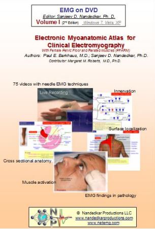
EMG/NCS Online Series: Volume I: Electronic Myoanatomic Atlas for Clinical Electromyography 2nd Edition 2020
$16
EMG/NCS Online Series: Volume I: Electronic Myoanatomic Atlas for Clinical Electromyography 2nd Edition 2020
YOU WILL GET THE COURSE VIA LIFETIME DOWNLOAD LINK (FAST SPEED) AFTER PAYMENT
Proper identification and activation of muscles is critical for assessment of needle EMG recordings. Dr Barkhaus methodically explains muscle identification using the anatomic landmarks and palpation. Upon identifying the site of needle insertion, live EMG signals are seen as the subject activates and relaxes the muscle. This collection of 74 videos covers the routinely tested muscles from upper limb, lower limb, neck, face and back (paraspinal). Dr Margret Roberts make an important contribution by discussing the pelvic floor muscles. Muscles used for chemo-denervation and some uncommon muscles are also included. The pictures show surface and cross sectional anatomy. A textbook in electronic format is also included for those who like printed copies.
Topics And Speakers:
– Cranial
– Distal Leg
– Foot
– Forearm – Extensors
– Forearm – Flexors
– Forearm – Rotators
– Hand
– Pelvic Floor and Related Muscles
– Pelvis
– Proximal Arm
– Shoulder
– Spinal
– Thigh
| Vendor |
|---|
Related products